 |
The Georgia Mineral Society, Inc. 4138 Steve Reynolds Boulevard Norcross, GA 30093-3059 |
GMS Field Trip
If you have any questions about field trips send email toGMS Field Trip
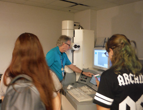
Georgia State University, Atlanta, GA
Saturday, August 5, 2017
Once again, Dr. Robert Simmons invited GMS members to tour the Imaging Core Facility at Georgia State University. Our tour started with laser confocal microscopes that are used for 3d imaging. Then Dr. Simmons explained brightfield microscopy where a specimen is illuminated from below. Next he demonstrated a transmission electron microscope (TEM). He loaded a specimen into the TEM chamber and focused on a bacteriophage, a virus that can be used to kill bacteria. We could see the capsid (head) and details of the tail. The core of the tail was visible and Dr. Simmons explained how the bacteriophage can use its tail to penetrate a bacterium’s cell wall and inject its DNA into the cell.
Then we saw an optical microscope, the sort of microscope you would use to look at minerals. That microscope was fitted with a camera with output to a computer. After looking at some dirt, we looked at a piece of a sock under normal light, then through a filter so we could examine it for fluorescence.
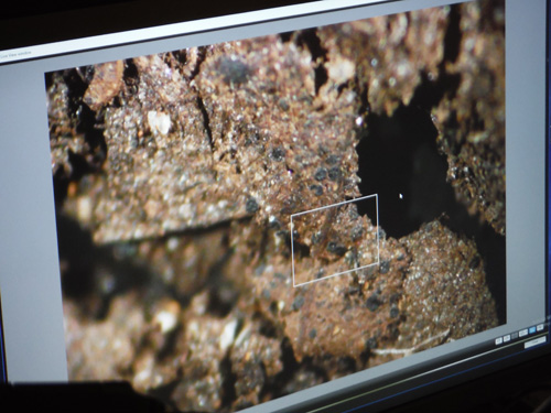
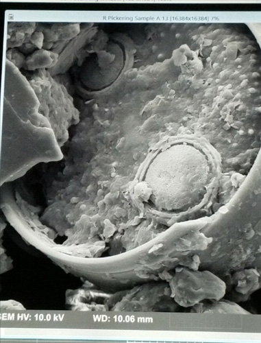
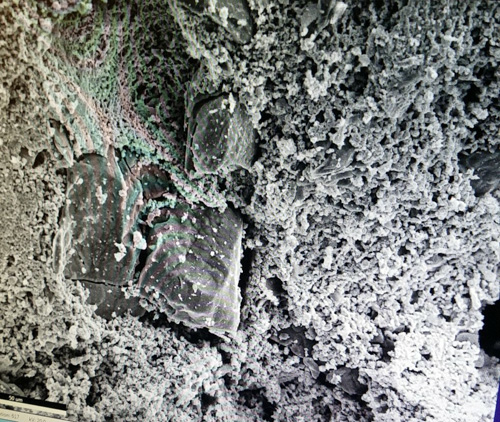
Next, members showed the samples we brought for the scanning electron microscope (SEM). Dr. Simmons selected a specimen of sand from Indonesia that I brought and a sample of the contents of a hollow sandstone concretion aka “indian paint pot” that Cameron and Cristina Clines brought. He prepped our samples with carbon to reduce static, then loaded them plus a pre-prepped diatom sample into the SEM. The first sample we looked at was from the paint pot. Though it looked like very fine powder to the naked eye, when viewed through the SEM we could see tiny ‘rocks”. Dr. Simmons ran x-ray analysis and we could see very clearly that the particles were primarily iron. The Indonesian sample was harder to work with as it still had some static, but we did get to see a few particles and Dr. Simmons will analyze it more when time allows. The last specimen we saw was the diatoms. There were different kinds of diatoms in the sample and they were all incredibly detailed. Dr. Simmons showed us images of other diatoms and fragment of cloth from a mummy!
Again, we cannot thank Dr. Simmons enough for inviting us to his lab and for taking time from his busy schedule to teach us about multiple types of microscopy. As always, the tour was informative, fascinating, and fun!
Lori Carter
On behalf of Charles Carter, GMS Field Trip Chair
e-mail:
Photo by Lori Carter
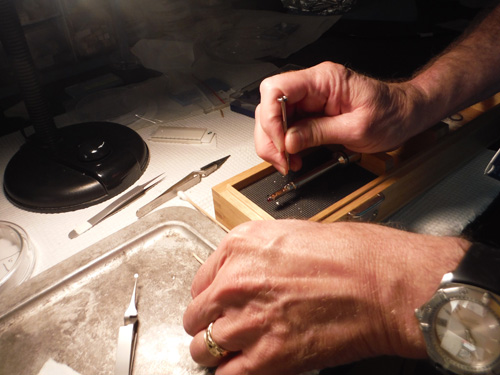
Dr. Simmons prepares a sample for the transmission electron microscope (TEM)
Photo by Lori Carter
Transmission electron microscope (TEM)
Photo by Cristina Clines
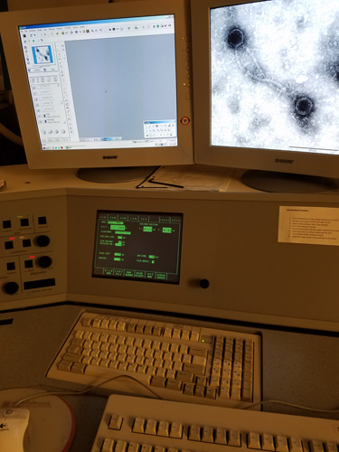
Using the TEM to see a bacteriophage
Photo by Cristina Clines
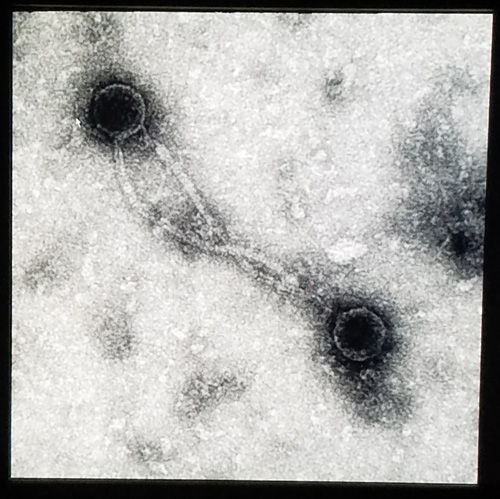
Close-up of the bacteriophage
Photo by Lori Carter
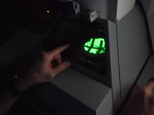
Another view of the TEM
Photo by Lori Carter
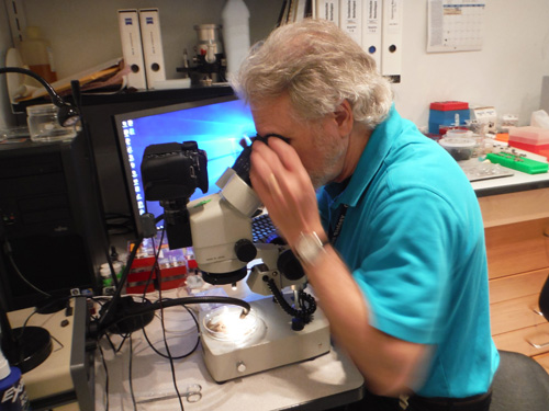
Dr. Simmons getting a sample ready under an optical microscope
Photos by Lori Carter
We looked at some dirt with the optical microscope...
Photo by Lori Carter

...then we looked at an old sock with the optical microscope with a filter for fluorescence
Photo by Lori Carter
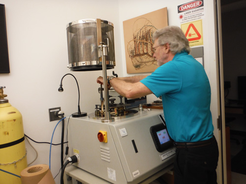
Dr. Simmons had to prep some samples with carbon to reduce static
Photo by Lori Carter
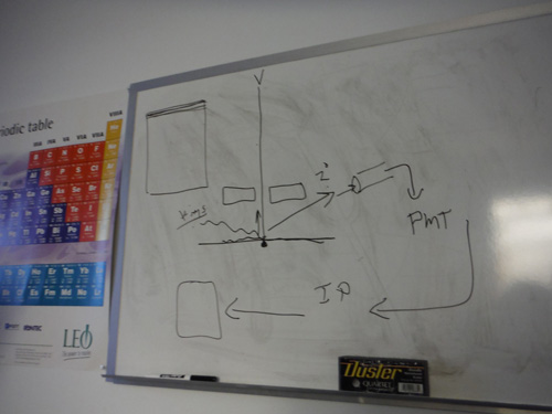
Dr. Simmons drew a diagram of where the light is directed in the scanning electron microscope
Photos by Cristina Clines
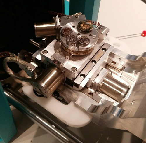
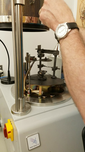
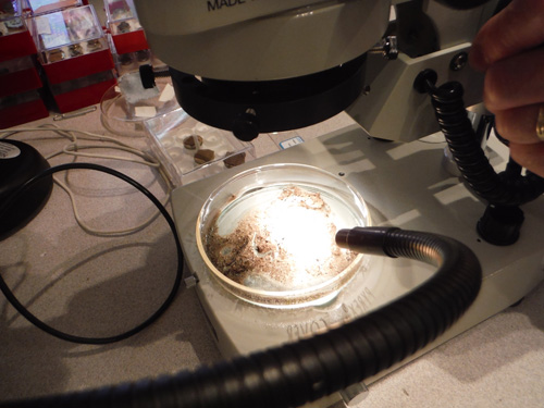
Getting the samples into the scanning electron microscope (SEM) chamber
Photos by Cristina Clines
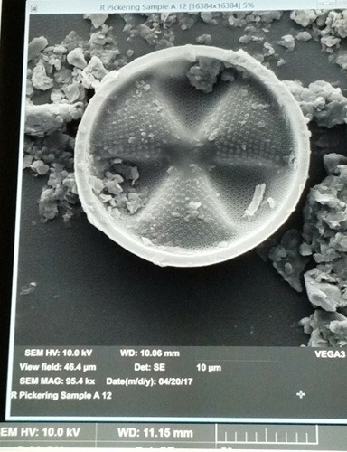
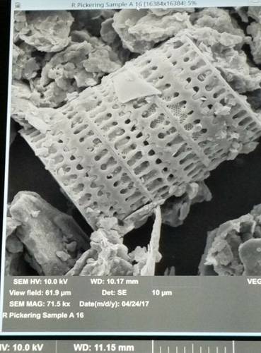

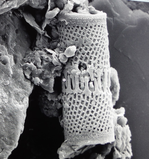
Some diatoms imaged with the SEM
Photo by Cristina Clines
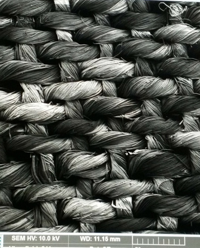
Fabric from a mummy!
Check back for images of the field trip specimens
after Dr. Simmons has time to analyze them
Click below for field trip policies

Copyright © Georgia Mineral Society, Inc.