GMS Field Trip
If you have any questions about field trips send email toGMS Field Trip
Georgia State University, Atlanta, GA
Saturday, February 11, 2017
Dr. Robert Simmons invited GMS members to visit the Imaging Core Facility at Georgia State University. We toured the lab where we got to see various microscopes in action. The first microscope on the tour was a brightfield/fluorescence microscope. I am sure it must be more cooperative when only Dr. Simmons sits quietly before it than when a group of ebullient field trip people are gathered around it, but Dr. Simmons was still able to show us some stunning images of a mineral sample.
Next, we saw a transmission electron microscope. The TEM was much more cooperative so we saw some fascinating images of a bacteria-defeating virus. We also got to see how a specimen is prepped for the microscope. Then we looked at some cloth under a stereo microscope via a DSLR camera outputting images to a computer. We could see details of the fiber all the way down to some aspergillus niger mold spores.
The last microscope on the tour was the scanning electron microscope. Field trip participants brought in several samples to view, and three were chosen -- a mineral in need of identification, some diatomaceous earth, and a clay sample that hopefully contained some phytoliths [Quiz: You look it up -- Tips and Trips editor]. The diatomaceous earth and clay samples had to be coated with gold for better imaging. While we waited for those, we got to see an already prepped sample of diatoms that Dr. Simmons collected from a river in Georgia. First we saw a strand of algae, then closer to see a chain of diatoms, then even closer to see a single diatom, where we could see a bacterium attached. So cool!
Charles Carter brought in some crystals from Arkansas. With the SEM, Dr. Simmons selected a couple of spots on a crystal. Then using X-ray microanalysis, he showed us the elements detected at the selected areas of the specimen. Charles hoped it was celestite, but the lack of strontium and sulfur confirmed it was not so we concluded it was calcite. I brought in some diatomaceous earth. The diatoms in that sample were fossilized, so it was interesting to see those compared to the "fresh" diatoms Dr. Simmons collected. Next we looked at Bill Montante's clay. Though we did not find any phytoliths, we did find some fossilized pine pollen -- wow!
To say it was a fascinating, thoroughly enjoyable trip is a woeful understatement. For Dr. Simmons, it was simply a demonstration of some of the tools of his profession. But for us, it was an extraordinary opportunity to get a tiny glimpse into a tiny universe. We can't thank Dr. Simmons enough for inviting GMS to his lab and teaching us so much about microscopy!
Lori Carter
On behalf of Charles Carter, GMS Field Trip Chair
e-mail:
Photo by Lori Carter
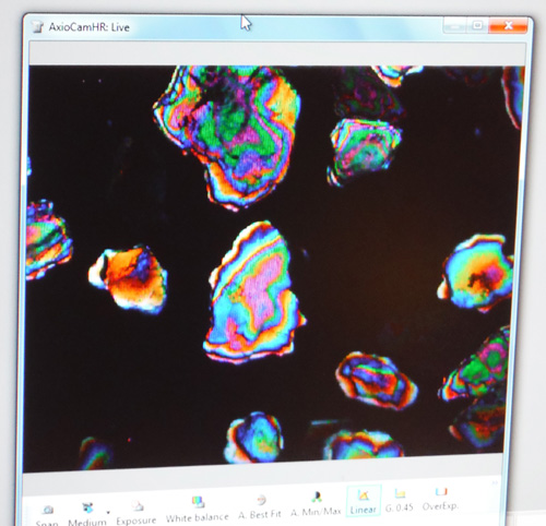
Brightfield/fluorescence microscope image of a mineral sample
Photo by Lori Carter
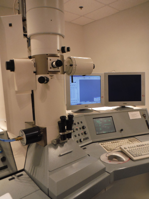
Transmission electron microscope (TEM)
Photo by Lori Carter
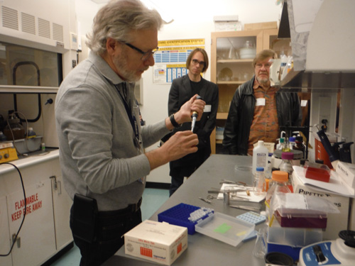
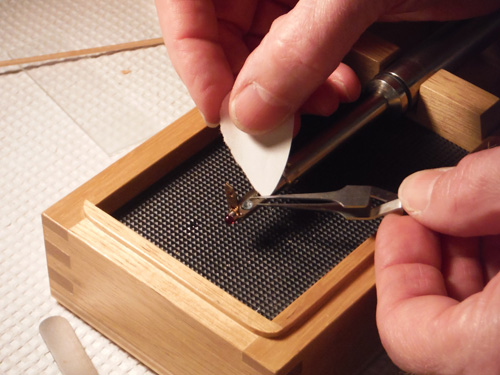
Dr. Simmons prepping a sample for the TEM
Photo by Lori Carter
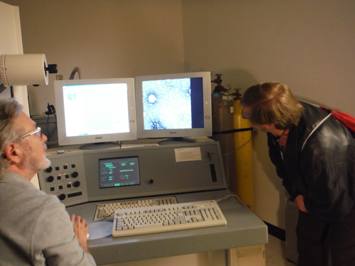
Everyone had a chance to get a good look at the TEM images
Photo by Lori Carter
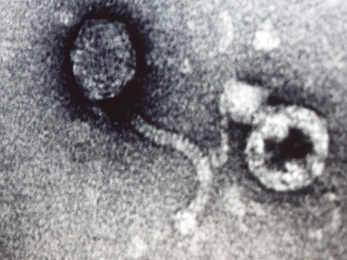
A close look at a virus with the TEM
Photo by Lori Carter
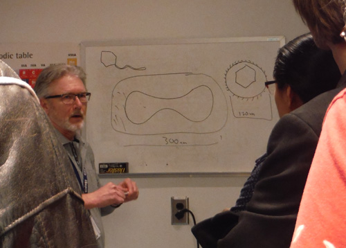
Dr. Simmons explained what we were seeing with the TEM
Photo by Lori Carter
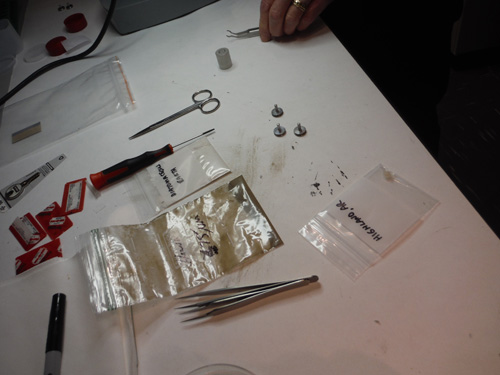
Field trip participants brought samples for the scanning electron microscope
Photo by Lori Carter
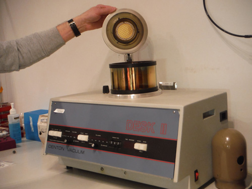
Some samples have to be coated with gold to get a good image with the SEM
Photo by Lori Carter
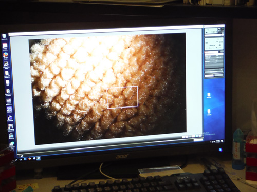
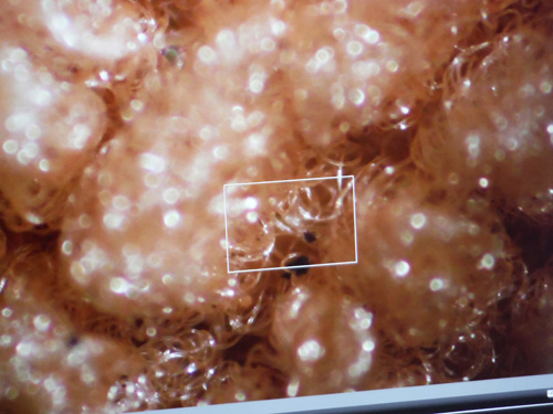
Fabric sample with Aspergillus niger mold
Photo by Lori Carter
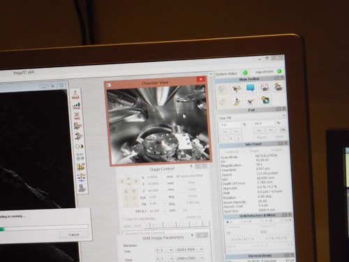
Live view of the SEM specimen chamber
Photos by Lori Carter
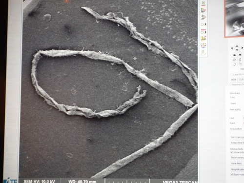
Algae strand
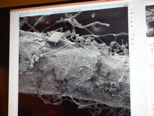
Closer view of algae showing diatoms
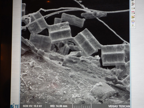
Closer view of diatoms
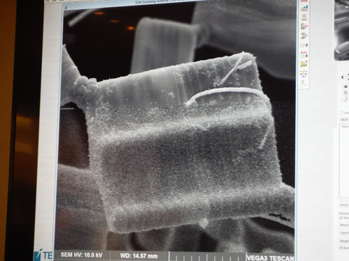
Single diatom with bacterium. Notice slower scan producing sharper image at the top
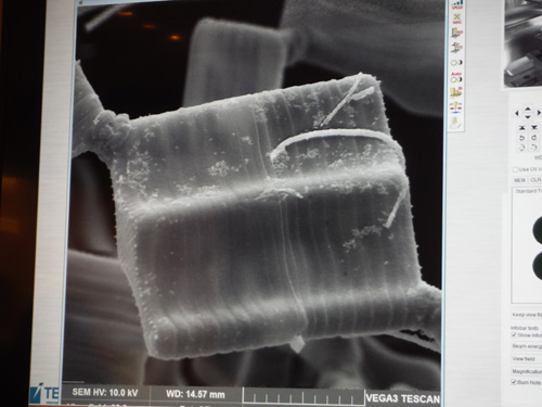
Same image as above after slower scan. Note fine details now visible.
Photo by Lori Carter
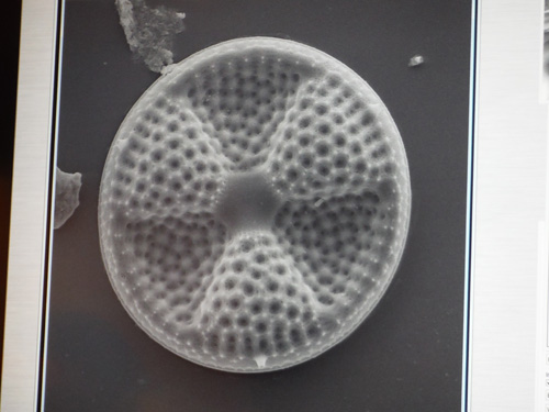
Another diatom
Photos by Lori Carter, SEM image by Dr. Simmons
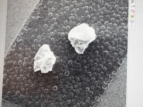
Unidentified mineral

SEM image. Note the 2 blue spots selected for analysis
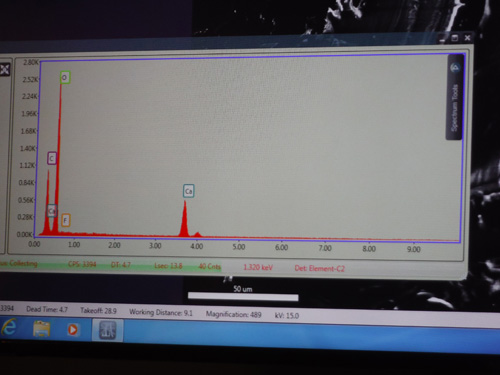
X-ray microanalysis results
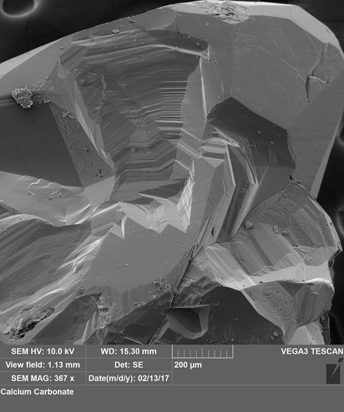
Another SEM image of the specimen
Images by Dr. Robert Simmons
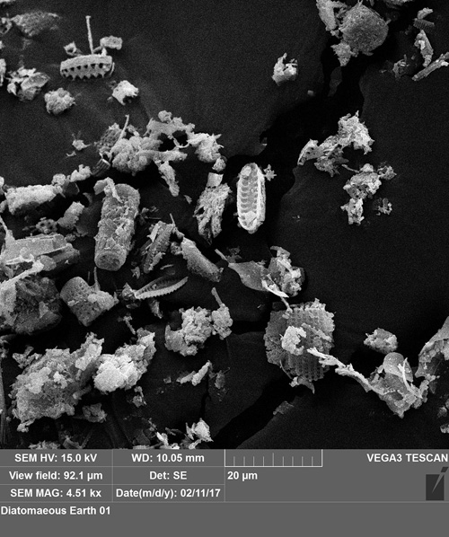
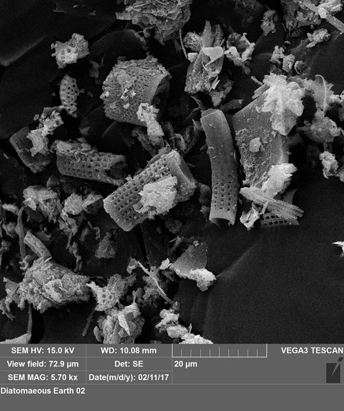
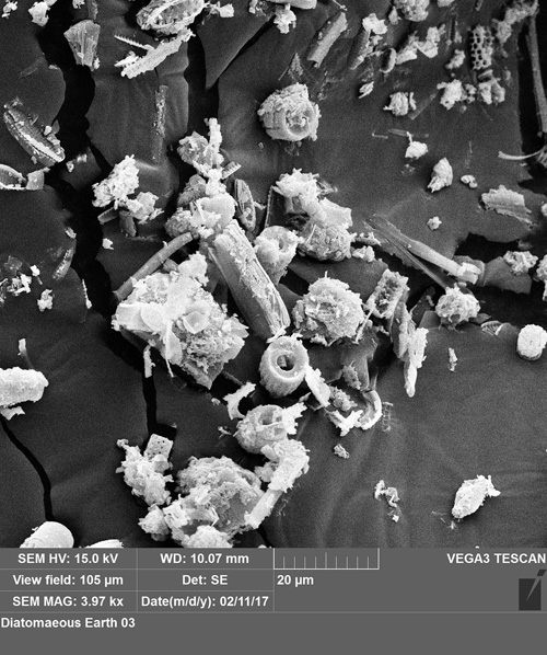
SEM images of the diatomaceous earth
Image by Dr. Robert Simmons
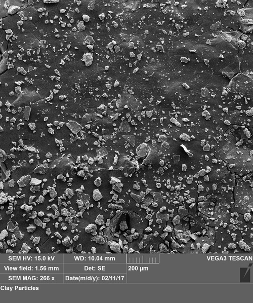
Bill Montante's clay sample image from the SEM
Images by Dr. Robert Simmons
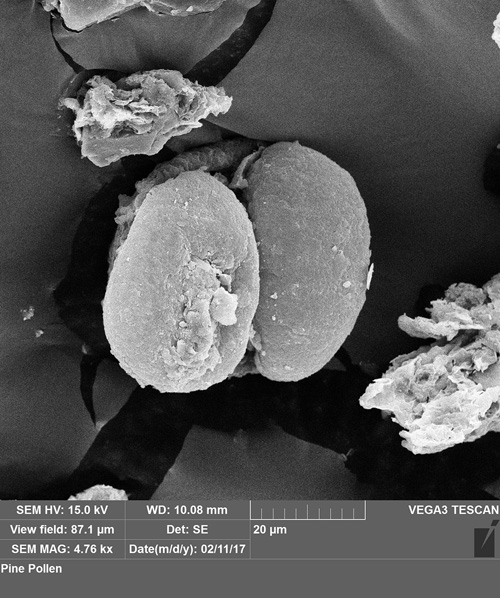
Fossilized pine pollen in the clay sample!
Click below for field trip policies

Copyright © Georgia Mineral Society, Inc.
